Scientific Program

Conference Series Ltd invites all the participants across the globe to attend 25th American Dental Research & Future Dentistry Montreal, Canada.

Day 2 :

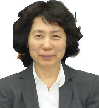
Conference Series Future Dentistry 2019 International Conference Keynote Speaker Jiin-Huey Chern Lin photo
Biography:

Jiin-Huey Chern Lin received her Ph.D. from Northwestern University Dept. of Biological Materials (Chicago, Illinois). She is a Professor of Dept of Mater Sci & Eng and Director of Center for Biomaterials Research, National Cheng-Kung University (Tainan, Taiwan). She also serves as Technology Founder/VP/CTO of Joy Medical Devices Corp. (Kaohsiung, Taiwan). She has published 100+ SCI papers along with
70+ US patents and 100+ non-US patents.

Abstract:

A series of synthetic, inorganic, osteoconductive/ osteoinductive and fully resorbable Ca/P/S-based bone substitute devices have been developed by a joint research project of National Cheng-Kung University and Joy Medical Devices Corporation of Taiwan. The granular type product (Ezechbone® Granule) is a highly porous material characterized by its non-inflammation, high resorption, and fast healing capability. The cement type product (Ezechbone® Cement) is featured by its non-inflammatory, injectable and non dispersive behavior in blood/body fluid in paste form. The safety and efficacy of these devices have been confirmed by a series of chemical/ physical characterization and biocompatibility tests. The animal models for implantation tests include the SD rat femur body, New Zealand white rabbit femur condyle and mandible, Lanyu pig mandible and osteoporotic goat spine. The rabbit condyle implantation/histopathologic examination indicates that the implants of both types are always intimately integrated with the surrounding bone substantially without a foreign body or other undesirable tissue reactions. The porous Ezechbone® Granule outperforms autologous bone by its less inflammatory/ irritative tissue response and higher new bone formation rate, especially at the critical early stage post-implantation. Clinical case reports covered in this presentation include dental ridge augmentation, sinus lift, sternum closure, frontal bone augmentation and treatment of various types of fractures.

Conference Series Future Dentistry 2019 International Conference Keynote Speaker Jimmy Kayastha photo
Biography:

Jimmy Kayastha, is a Consultant in Oral and Maxillofacial Medicine and Surgery at Dental Health Solutions Inc., San Francisco, California.

Abstract:

Background: The purpose of this study was to assess the effect of free-flap reconstruction of patients with advancedstage IV oral squamous cell carcinoma following ablative tumor resection. The purpose of this study is to elucidate the reconstructive indications of
the use of simultaneous double free flaps in head and neck oncological surgery.
 
Method: The study was based on a restrospective cohort of 76 patients with pathological stage IV OSCC patients (without distant metastasis) treated by tumor ablation with free flap reconstruction. Of the 76 patients, 49 (Group 1, Test) underwent surgical reconstruction with microvascular tissue transfer and in 27 (Group 2, Control) only local or regional flaps were used. Fibula osteo-cutaneous free flap was used in association with forearm free flap in 18 cases, fibula osseous-forearm in 7 cases, fibula osseous-rectus abdominis in 1 case, iliac crest-forearm in 1 case. Forearm free flap was used for intra-oral reconstruction in all cases. We compared patient survival and cancer recurrence rates between these two groups.
 
Result: Despite the unfavorably expected prognosis in group 1, both positive margin rate (12.2% in Group 1 versus 21.5% in Group 2, P = 0.112) and cancer recurrence rate (26.6% in Group 1 versus 28.3% in Group 2; P = 0.671) were not significantly different between the two groups. At the end of the follow-up period, 23 (47%) and 33 (67.3%) patients had died of oral squamous cell carcinoma in the microvascular reconstructive and control group, respectively. In the free-flap group, the mean and median survival time was 54 months. In the locoregional flap group, the mean and median survival time was 51 months respectively. No difference was seen in the survival time between the free-flap and local flap groups (P = .2). Univariate Kaplan Meier analysis revealed that positive surgical margins were significantly associated with shortened survival in the free-flap group and that recurrence was significant in both reconstructive groups. On multivariate Cox regression analysis, the status of the resection margin (P = .05) and tumor recurrence (P < .0004) showed a significant relationship with survival.
 
Conclusion: Patients with free-flap reconstruction of surgically created defects after oral cancer resection showed a trend toward better 5-year survival. Simultaneous free flap reconstruction, in massive oromandibular defects, represents in some selected patients, a good choice to achieve satisfactory aesthetic and functional results.

Keynote Forum

Fatma Elhendawy

Tanta University, Egypt

Keynote: Pediatric full mouth rehab under general anesthesia
Biography:

Fatma Elhendawy is currently working as an editorial member of Tanta Dental Journal and professor.

Abstract:

General anesthesia is an important technique to manage the child behavior. It is a controlled state of unconsciousness in which there is a loss of protective reflexes, including the ability to m maintain an airway independently and to respond appropriately to physical stimulation or verbal command. Indications and contra indications of general anesthesia must be considered according to the classification of American society of anesthesiologist. The anatomic and physiologic differences between child and adult also are very important issues during the application of general anesthesia .Pre-operative evaluation and preparation of the patient with the operative procedures will be included together with postoperative instructions and Complications of general anesthesia.

  • Prosthodontics | Dental Materials | Digital Dentistry | Dental SEO | Dental Marketing | Dental Surgery | Oral Microbiology and Pathology | Endodontics | Dental Nursing | Oral Implantology
Location: Conference Hall
Speaker

Chair

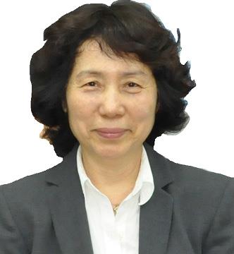
Jiin-Huey Chern Lin,

National Cheng-Kung University, Taiwan

Co-Chair

Fatma Elhendawy

Tanta University, Egypt

Biography:

Marwa Baraka is working in Tanta University, Egypt


Abstract:

Introduction: Various forms of malocclusion are usually encountered in non syndromic cleft lip and palate, mostly affecting maxillary dental arch.

Objective: To assess the dental arch parameters in surgically treated unilateral cleft lip and palate Egyptian children with mixed dentition and compare them with those of comparable healthy non-cleft children.

Material and methods: Comparative cross-sectional study design was used. Twentysix non-syndromic children with repaired unilateral cleft lip and palate (UCLP), aged 6-9 years, were compared to twentysix healthy non-cleft children (control group) recruited from Faculty of Dentistry, Alexandria University. Both groups were divided into two age groups; 6-7 years and 8-9 years. For each subject, dental arch parameters were measured using dental study models.

Results: Mean maxillary arch depth and inter-canine arch width were significantly smaller in UCLP children than in non-cleft children in the age groups 6-7 and 8-9 years. Mean inter-molar arch width was not significantly narrower in UCLP children than in non-cleft children. Mean mandibular arch dimensions of UCLP children did not differ significantly from those of non-cleft children. The mean Goslon Yardstick score was 3. Conclusions: Children with UCLP, aged 6-9 year old, had significant reduction in mean maxillary arch dimensions compared to healthy matching non-cleft children except for inter molar arch width which showed no significant reduction. The average dental arch relationship in surgicallyrepaired UCLP children was fair according to Goslon Yardstick index.

Biography:

LOGHMAN REZAEISOUFI is Associate Professor who is working as a Faculty member at the Department of Restorative Dentistry, Dental Research Center, Dental Faculty, Hamadan University of Medical Sciences, Hamadan, Iran He has completed his DDS degree at the age of 25 years from Tehran University of Medical Sciences and also has continued Specilized Restorative (operative) Dentistry felowship in Mashahd University and Laser felloship course at the Achen University. He has published more than 30 papers in reputed journals and has been serving as an editorial board member of repute.

Abstract:

Nowadays, studies show that laser irradiation can enhance the adhesion of
bonding agents to the dentin. Therefore, the present study was set to find an appropriate protocol for the use of Nd:YAG, Er:YAG, and 940 nm diode lasers
during the application of the 6th generation of adhesives for dental restorations. A total number of 100 healthy extracted teeth were distributed in the ten groups. Control group(C) was prepared according to the manufacturer’s guideline. Each of the remaining 9 groups were treated with Er:YAG, Nd:YAG
and 940 nm diode lasers in following stages: Before applying primer (Er BP,Nd-BP,D-BP), After applying primer (Er-AP,Nd- AP,D-AP) and after applying bonding (Er-AB,Nd-AB,D-AB. The strength of microtensile bonding was measured using the universal testing machine. The data were analyzed using one-way ANOVA followed by post hoc Tukey test (p<0.05). The results of the means and standard deviation of the groups were as follow: C=30.09±4.21,
Er-BP=18.83±4.21,
Er-AP=14.43±3.12,
Er-AB=19.67±4.96,
Nd-BP=20.35±5.55,
Nd-AP=39.85±4.13,
Nd-AB=18.16±3.36,
D-BP=26.74±5.05,
D-AP=28.11±5.12,
D-AB=37.28±5.61.
The means of two groups: Nd:AP and D-AB were significantly higher than control groups. (p<0.05) D-BP and D-AP had no siginificant difference in
comparison with control group while the remaining groups had significantly lower values (P<0.05). It was demonstrated by the present study that implementing the Nd: YAG laser system after the primer and also implementing the 940nm diode laser after the bonding significantly improved the bond strength.

Biography:

Argishti Hayrapetyan completed a Bachelor of Science in 2011 and pursued to obtaining his Master of Science degree in 2013 from Yerevan State Medical University. Thereafter, the General Family Dentistry residency was completed in 2015. He is a member of the Armenian Dental Association, attended University Hospital of Olomouc, Czech Republic, Division of Dentistry and Oral Surgery for professional exchange in complex dental restorations. He is currently in the field of restorative dentistry, specializing in diagnosis and management of diseases of the teeth and their supporting structures and the
rehabilitation of the dentition to functional and aesthetic requirements of the individuals at “Drampyan Clinic” in Yerevan, Armenia.

Abstract:

Veneer restorations are one of the most conservative and aesthetic techniques
applied as treatment of unaesthetic anterior teeth as they avoid aggressive dental preparation; thus, maintaining tooth structure. The continued development of materials offers clinicians many options for creating highly aesthetic and functional laminate veneers. Correction of minor malformations of anterior teeth with minimal removal of the remaining tooth structure, correction of crown length and width, diastema closure and masking, as well as some intrinsic discoloration of anterior teeth are examples of solutions that can be gained with using porcelain veneers. Initially, laminates were placed on the
unprepared tooth surface. Although there is as yet no consensus as to whether or not teeth should be prepared for laminate veneers, currently, more  conservative preparations have been advocated. Because of their aesthetic appeal, biocompatibility, and adherence to the physiology of minimalinvasive
dentistry, porcelain laminate veneers have now become a restoration of choice.
Today, limited controversial information is available regarding the influence of preparation design on the longevity of laminate restorations. The purpose of this presentation is to attempt to review the different incisal preparation designs of porcelain laminate veneers for anterior teeth. A further objective is to understand which is the most successful design in terms of preparation.

Biography:

Soliman Ouda is working in King Abdulaziz University, Saudi Arabia

Abstract:

Objective: To study and compare the efficacy of Vizilite®Plus and computer-assisted brush biopsy (Oral CDx), a non-invasive adjuvant diagnostic aids, in the detection and assessment of oral potentially malignant, and/or malignant lesions.
 
Study Design: This is a prospective study that included a total of 500 patients referred to Oral Medicine clinics of King Abdulaziz University Faculty of
Dentistry (KAUFD), and regional Ministry of Health (MOH) hospitals, presenting with oral mucosal lesions over a 24-month period which were referred for further management. All cases were examined with Vizilite®Plus.
Cases with positive results were subjected to Oral CDx. Positive or atypical Oral CDx lesions, as well as negative Oral CDx lesions with clinical suspicion of precancer/ malignancy, were scalpel biopsied.
 
Results: The sensitivity of Vizilite®Plus in detecting dysplasia/malignancy was
100% whereas the specificity was 30.3%. The positive predictive value was 19.29%, whereas the negative predictive value was 100%. The positive
likelihood ratio was 1.43 and the negative likelihood ratio was 0. The sensitivity of Oral CDx in detecting dysplasia/malignancy was 97.4%, whereas
the specificity was 78.94%. The positive predictive value was 52.44% whereas the negative predictive value was 99.22%. The positive likelihood ratio was 4.6
and the negative likelihood ratio was 0.03.
 
Conclusions: Because of its low positive and negative predictive values, we do not advocate Vizilite®Plus as a useful adjunctive tool for the diagnosis of oral cancer. Oral CDx is more promising and can be utilized as an aid for the early diagnosis of suspicious oral lesions with limitations.

Biography:

Abstract:

The term “diabetes mellitus” denotes a number of diseases marked by high levels of glucose in the blood and abnormalities of fat, carbohydrate, and protein metabolism. A group of oral diseases and disorders has been associated with diabetes mellitus, and periodontitis has been identified as a probable risk
factor for poor metabolic control in subjects with diabetes.
 
Methods: The researcher reviewed the literature to characterize oral conditions that are hit by diabetes mellitus. She also scrutinized the literature related to periodontitis as a modifier of glycemic control.
 
Results: Despite the fact that a group of oral diseases and disorders has been linked to diabetes mellitus, the data braces the fact that periodontitis is a  complication of diabetes. Patients with constant and poorly controlled diabetes are at higher risk of developing oral candidiasis, and the evidence shows that periodontitis is a risk factor for poor glycemic control and the development of other clinical complications of diabetes. Evidence suggests that periodontal changes are the first clinical demonstration of diabetes.

Biography:

Mehdi Kadkhodazadeh has completed his Doctor of Dentistry from Shahid Beheshti University of Medical Science and Periodontology from the Mashhad University of Medical Science. She is the Director of Periodontology. She has published more than 40 papers in journals.

Abstract:

In inflammatory diseases such as peri-implantitis (PI) and chronic periodontitis (CP), both adaptive and innate immunity play a part. Natural resistanceassociated macrophage protein 1(Nramp1) has considerable effects on macrophage function (phagocytosis) and host innate immune response against infections. The present study was to investigate the relationship of Nramp1 gene polymorphisms with PI and CP in an Iranian population. In this crosssectional study 79 patients with CP, 38 patients with PI and 84 healthy controls presenting to the Periodontology Department of Shahid Beheshti University of Medical Sciences were enrolled. DNA was extracted from fresh blood samples of arm vein of participants and transferred to the KBiosience Institute (United Kingdom) for genotyping. χ2 and Fisher’s exact tests were
used by SPSS software v.19 for statistical analyses. Significant differences were detected in the distribution of genotypes between control and CP groups
both for rs17235409 and rs2276631 polymorphisms (P:0.044 and P:0.028
respectively). Distribution of genotypes differed insignificantly in comparison of PI and control groups for rs2276631 (P:0.623) and either rs17235409 (P:1)
polymorphisms. Based on our results, we conclude that the presence of G allele in both rs2276631 and rs17235409 location may be a protective factor against CP. More studies with a larger sample size in different populations are required for confirming Nramp1 as a genetic determinant in periodontal disorders.

Biography:

Abeer Ahmed has completed her M.Sc. from King Saud University in 2017. Currently, she is studying the doctorate program from King Saud University College of Dentistry. In addition, she is a researcher in King Saud University at Periodontology and Community Dentistry Department.

Abstract:

A gummy smile is an excessive display of the gingiva or imbalance in the gingival-tooth ratio. It has different etiological factors such as vertical maxillary excess, short upper lip and altered passive eruption which cause a major aesthetic concern for many patients that may be reflected in their social or behavioral daily life, thus several techniques have been developed for its treatment. Treatment varies according to the etiological factors,
perception, and expectations of the patients like orthodontics, orthognathic surgery, cosmetic surgery, and periodontal surgery (including gingivectomy,
apically repositioned flap and lip repositioning technique). Lip repositioning surgery, chosen mainly in the treatment of hypermobile upper lip and excess
gingival display, consists of the removal of a stripe of mucosa from the maxillary vestibule and suturing the lip mucosa with the mucogingival junction. It is well tolerated by patients and results are irreversible with a very high rate of satisfaction. This report will present the results of clinical cases and the demonstration of the procedure step by step. Indications, objectives, limitations and patient’s outcome will be explained.

Biography:

Sung Ho Kim has completed his bachelor’s degree at the age of 25 years from Dankook University. He is currently a resident at the Department of Prosthodontics, Dankook University and studying for a master’s degree of prosthodontics. He participates in as a member of The Korean Academy of Prosthodontics (KAP) and The International Team for Implantology (ITI). His current research focuses on Ceramic Prosthesis, Digital Dental Technology, and Implant Restorative Dentistry. He is involved in basic and clinical research on Oral Implantology and Applied Digital Dental Technology.

Abstract:

The treatment of areas demanding esthetic requirements, such as maxillary anterior teeth, should take into account the achievement of a healthy, harmonious to the surrounding tissue, and an attractive smile line. In this case report, the smile line, soft tissue and hard tissue morphology, and the anatomy and proportion of the tooth must be considered. In patients with an unesthetic maxillary anterior ratio due to inadequate gingival contour and diastema caused by peg laterals in maxillary lateral incisors, it would be challenging to achieve
an esthetic restoration by orthodontic treatment alone. In such a case, after orthodontic treatment, gingivo-dentofacial compositional diagnosis and analysis, followed by gingivectomy and prosthodontic restoration are needed to improve the interdental mesial/
distal, width/length ratio to achieve a satisfactory aesthetic result. In addition, when improving the tooth proportion of peg laterals by prosthodontic treatment, Porcelain laminate veneer (PLV), which results in less tooth structure loss, reproduction of similar shade to that of the proximal tooth and high transparency, is recommended. This case report demonstrates esthetic improvements by prosthodontic restoration through systematic diagnosis and treatment procedure in patients with an unesthetic maxillary anterior
proportion after orthodontic treatment due to peg laterals in maxillary lateral incisors by
means of two female patients aged twenty years old.

Biography:

Marwa Baraka is currently working in Tanta University, Egypt

Abstract:

Introduction: Various forms of malocclusion are usually encountered in non-syndromic cleft lip and palate, mostly affecting maxillary dental arch. Objective: To assess the dental arch parameters in surgically treated unilateral cleft lip and palate Egyptian children with mixed dentition and compare them with those of comparable healthy non-cleft children.
 
Material and methods: Comparative cross-sectional study design was used. Twentysix non-syndromic children with repaired unilateral cleft lip and palate (UCLP), aged 6-9 years, were compared to twentysix healthy non-cleft children (control group) recruited from Faculty of Dentistry, Alexandria University. Both groups were divided into two age groups; 6-7 years and 8-9 years. For each subject, dental arch parameters were measured using dental
study models.
 
Results: Mean maxillary arch depth and inter-canine arch width were significantly smaller in UCLP children than in non-cleft children in the age groups 6-7 and 8-9 years. Mean inter-molar arch width was not significantly
narrower in UCLP children than in non-cleft children. Mean mandibular arch dimensions of UCLP children did not differ significantly from those of non-cleft children. The mean Goslon Yardstick score was 3.
 
Conclusions: Children with UCLP, aged 6-9 year old, had significant reduction in mean maxillary arch
dimensions compared to healthy matching non-cleft children except for inter-molar arch width which showed no significant reduction. The average dental arch relationship in surgicallyrepaired UCLP children was fair according to Goslon Yardstick index.